Why are crystals used for electron diffraction?
Why are crystals used for electron diffraction?
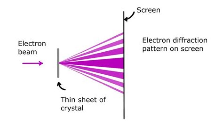
The periodic structure of a crystalline solid acts as a diffraction grating, scattering the electrons in a predictable manner. Working back from the observed diffraction pattern, it may be possible to deduce the structure of the crystal producing the diffraction pattern.
What can electron microscopy tell us beyond crystal structures?
When individual particles are examined using electron microscopy, much more information beyond average crystal structures can be obtained, including particle size and shape, crystal orientation, defects, crystallinity, surface structures, superstructures, etc.
What does electron microscopy use?
The electron microscope uses a beam of electrons and their wave-like characteristics to magnify an object’s image, unlike the optical microscope that uses visible light to magnify images.
How does electron crystallography work?
Electron crystallography is similar to X-ray crystallography in that a protein crystal scatters a beam to produce a diffraction pattern. However, the interactions between the electrons in the beam and the crystal are much stronger than those between the X-ray photons and the crystal.
How diffraction is used in electron microscope?
The use of electromagnetic lenses allows diffracted electrons to be focused into a regular arrangement of diffraction spots that are projected and recorded as the electron diffraction pattern. If the transmitted and the diffracted beams interfere on the image plane, a magnified image of the sample can be observed.
What is electron diffraction technique?
Electron diffraction is a technique that allows determination of the crystal structure of materials. When the electron beam is projected onto a specimen, its crystal lattice acts as a diffraction grating, scattering the electrons in a predictable manner, and resulting in a diffraction pattern.
What does TEM stand for?
TEM
| Acronym | Definition |
|---|---|
| TEM | Transmission Electron Microscope |
| TEM | Transmission Electron Microscopy |
| TEM | Telecom Expense Management |
| TEM | Työ-Ja Elinkeinoministeriön (Finnish: Labor and Industries Ministry) |
Can Tem be used to measure crystallinity index?
Q: Can TEM be used to measure crystallinity index as XRD? A: In a single crystal sample, we get well-resolved diffraction patterns that we can use to measure lattice parameters and similar aspects of crystallinity.
Can an Electron Microscope see a virus?
Viruses are very small and most of them can be seen only by TEM (transmission electron microscopy). TEM has therefore made a major contribution to virology, including the discovery of many viruses, the diagnosis of various viral infections and fundamental investigations of virus-host cell interactions.
Are electron microscope images real?
The image below on the right is the real image taken by a transmission electron microscope. You can see the scale bar (100 nm) below with a magnification 150,000x. In addition, the EM images are black and white. Therefore, the right image is the real image via an electron microscope.
How does cryo electron microscopy work?
The technique involves flash-freezing solutions of proteins or other biomolecules and then bombarding them with electrons to produce microscope images of individual molecules. These are used to reconstruct the 3D shape, or structure, of the molecule.