The cingulum refers to the portion of the teeth (anterior teeth (incisors and canines)), occurring on the lingual or palatal aspects, that forms a convex protuberance at the cervical third of the anatomic crown. It represents the lingual or palatal developmental lobe of these teeth..
Accordingly, where is the Cingulum located?
The cingulum is described from various brain images as a C shaped structure within the brain that wraps around the frontal lobe to the temporal lobe right above the corpus callosum. It is located beneath the cingulate gyrus within the medial surface of the brain therefore encircling the entire brain.
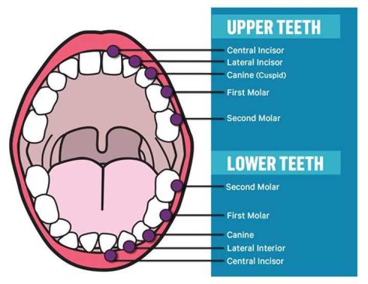
Also, what are the 5 surfaces of a tooth? The crown of each tooth has 5 surfaces, as follows:
- Buccal (facing the cheek or lip)
- Lingual (facing the tongue)
- Mesial (between the teeth)
- Distal (between the teeth)
- Chewing (occlusal for molars and premolars, incisal for incisors and canines)
Secondly, which tooth has the most prominent Cingulum?
On the palatal aspect, the marginal ridges and cingulum are more prominent. It has the most cervically located contact area of any incisor. Next to third molars, maxillary lateral incisors are the teeth that show most variation in crown size, shape and form (see Figure 8).
Why do my teeth have ridges?
These ridges/grooves are known as “Mamelons”. They appear on newly emerged adult teeth because of the way they develop. Mamelons do not typically last long since they are uneven and thin. Those who still have mamelons and do not like the appearance of them, can have them smoothed out by their dentist.
Related Question Answers
What is the long axis of the tooth?
The long axis of a tooth is an imaginary line that goes through the crown and root around which the substance of a tooth is most symmetrically distributed. Any surface of a tooth that is parallel to the long axis is called an axial surface (for example, mesial, distal, facial, or lingual surfaces).What is the function of the cingulate cortex?
The cingulate cortex is usually considered part of the limbic lobe. It receives inputs from the thalamus and the neocortex, and projects to the entorhinal cortex via the cingulum. It is an integral part of the limbic system, which is involved with emotion formation and processing, learning, and memory.Which teeth have Imbrication lines?
Macroscopically, these lines can be seen on the labial surface or lip side of anterior or front teeth as horizontal lines on the tooth crown, also known as perikymata or "imbrication lines" . Evenly spaced Retzius lines indicate a 6- to 11-day cycle of enamel formation.Which tooth has the shortest root?
mandibular central incisor
Which tooth has the longest root?
mandibular canine
Which tooth is bilaterally symmetrical?
Bilateral symmetry in the maxillary incisor teeth is of significant importance in esthetic dentistry. In restorative dentistry, symmetry refers to the appearance of balance around the dental midline.Which tooth has the longest root in the maxillary arch?
canines
Which teeth are responsible for tearing food?
Incisors — the sharp, chisel-shaped front teeth (four upper, four lower) used for cutting food. Canines — sometimes called cuspids, these teeth are shaped like points (cusps) and are used for tearing food.How many central incisors are there?
There are 16 teeth in the maxilla and 16 in the mandible. In each arch there are two central incisors, two lateral incisors, two canines, four premolars, and six molars. The permanent central incisors, lateral incisors, first and second premolars replace the primary dentition.What is mesial side of tooth?
Occlusal – the top surface or chewing surface of the tooth. Mesial – this is a side surface of the tooth; the side that is closer to the front of the mouth. Distal – this is also a side surface of the tooth, the side that is closer to the back of the mouth.What is incisal surface of tooth?
Incisal – The biting edge of an anterior tooth. Lingual – The surface that faces the tongue. Mesial – The surface that is closest to the midline of the face. Occlusal – The chewing surface of posterior teeth.What is buccal side of tooth?
Buccal – The cheek-side of the tooth. This surface is also referred to as the facial surface when referring to the front teeth. Lingual – The part of the tooth that is closest to the tongue.What does Mo mean in dentistry?
This photo shows teeth #2-5 (Universal numbering system). Tooth #3, the upper right first molar, has an MO (mesial-occlusal) gold inlay. This molar is both posterior, as well as distal, to the premolars in front of it.Does a buccal filling hurt?
When a person has a cavity in their tooth, a dentist will probably recommend a filling. Fillings are safe and effective, but some people might experience discomfort or tooth sensitivity afterward. Most of the time, this sensitivity is normal and will resolve within a few days or weeks.Are teeth bones?
Teeth consist mostly of hard, inorganic minerals like calcium. They also contain nerves, blood vessels and specialized cells. But they are not bones. Teeth don't have the regenerative powers that bones do and can't grow back together if broken.What is the palatal surface?
Description. The palatal surface of a tooth is directed toward the palate, as opposed to buccal, labial or facial which refer to the side of a tooth adjacent to (or the direction towards) the inside of the cheek or lips, respectively. This term is strictly used in the maxilla.What is the anatomy of a tooth?
Your teeth are composed of four dental tissues. Three of them—enamel, dentin and cementum—are hard tissues. The fourth tissue—pulp, or the center of the tooth that contains nerves, blood vessels and connective tissue—is a soft, or non-calcified, tissue.