What is the muscularis propria
There are usually two layers; the inner layer is circular, and the outer layer is longitudinal. These layers of smooth muscle are used for peristalsis (rhythmic waves of contraction), to move food down through the gut.
What is the muscularis propria made of?
It is composed of elastic fibers and 3–10 smooth muscle cells, generally arranged in an outer longitudinal and inner circular layer. Smooth muscle cells may radiate from the muscularis mucosa into the lamina propria and extend in the villi.
What is the difference between muscularis mucosa and muscularis propria?
The mucosa surrounds the lumen of the GI tract and consists of an epithelial cell layer supported by a thin layer of connective tissue known as the lamina propria. The muscularis mucosa is a thin layer of smooth muscle that supports the mucosa and provides it with the ability to move and fold.
What is the function of the Muscularis?
The muscularis externa is responsible for segmental contractions and peristaltic movement in the GI tract. These muscles cause food to move and churn together with digestive enzymes down the GI tract.What is lamina propria and muscularis mucosa?
The lamina propria is one of three layers which make up the mucosa, or mucous membrane. The lamina propria is a large layer of connective tissue which separates the innermost layer of epithelial cells from a layer of smooth muscle tissue called the muscularis mucosa.
What type of tissue is found in the muscularis?
The muscularis mucosae is a thin layer of smooth muscle at the boundary between mucosa and submucosa. It occurs throughout the GI tract from esophagus to rectum. It is thickest in esophagus, where it consists of relatively conspicuous bundles of longitudinal muscle fibers.
What does muscularis mucosa of stomach consist of?
In the stomach, muscularis mucosa is composed of two thin layers of smooth muscles arranged as inner circular and outer longitudinal layer. The muscularis externa is three layered thick as outer longitudinal, middle circular and inner oblique layer and are oriented more randomly than layered [2].
What protects the esophagus?
Non-keratinized stratified squamous epithelial tissue makes up the majority of the mucosa layer and provides protection to the esophagus from rough food particles and acid from the nearby stomach. Mucous glands in the mucosa produce mucus to lubricate the esophagus and help shield the mucosa from stomach acid.Is the Muscularis Mucosae part of the lamina propria?
The lamina muscularis mucosae (or muscularis mucosae) is a thin layer (lamina) of muscle of the gastrointestinal tract, located outside the lamina propria, and separating it from the submucosa.
In which layer is the lamina propria found?Lamina propriaFMA62517Anatomical terminology
Article first time published onWhere is the muscularis propria of the bladder?
The lamina propria (also called the submucosa) is a thin layer of connective tissue that surrounds the urothelium. It contains blood vessels, nerves and glands. The muscularis propria is the thick, outer muscle layer of the bladder.
What is the anterior region of small intestine called?
The small intestine has three distinct regions – the duodenum, jejunum, and ileum. The duodenum, the shortest, is where preparation for absorption through small finger-like protrusions called villi begins.
What is lamina propria in trachea?
The wall of the trachea is composed of a mucosa, submucosa, cartilaginous layer, and adventitia. The lamina propria of the mucosa contains many elastic fibers, lymphoid tissue in diffuse patches, and occasional small nodules. … This muscle is smooth muscle and attaches to the free ends of the cartilage rings.
What are the three layers of the lamina propria?
- The Vocalis Muscle (labeled above as the muscularis)
- The Lamina Propria (really 3 layers: deep, intermediate, and superficial)
- The epithelium or epithelial tissue.
Which of the following layers that contain muscularis mucosa?
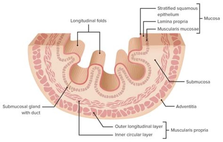
The GI tract contains four layers: the innermost layer is the mucosa, underneath this is the submucosa, followed by the muscularis propria and finally, the outermost layer – the adventitia.
Does the appendix have muscularis mucosa?
The appendix is completely invested by peritoneum, and has both an inner circumferential and a fully circumferential, outer longitudinal muscle layer of the muscularis propria. The mucosa of the appendix is colonic in type.
Is muscularis mucosa a muscle?
The muscularis mucosa is a thin layer of muscle whose contraction folds the mucosa to form ridges and valleys. Below the muscularis mucosa is the submucosa containing connective tissue, blood vessels, and nerves.
What cells are found in the crypts of Lieberkuhn?
The enteroendocrine cells are located within the Crypts of Lieberkuhn. They secrete hormones in response to various stimuli. There are four main classes of enteroendocrine cell, each with a different secretory product. These are I cells, S cells, K cells and enterochromaffin cells.
What does lamina propria look like?
What does the lamina propria look like under the microscope? The lamina propria is a very thin layer of tissue that can only be seen under the microscope. It is made up of long, thin supporting cells called fibroblasts, which make specialized matrix proteins that hold the tissue together.
What is lamina propria inflammation?
Chronic gastritis is a persistent inflammatory reaction in the gastric mucosa that is characterized by the accumulation of lymphocytes and plasma cells in the lamina propria. Chronic active gastritis implies that ongoing active inflammation is causing damage to epithelial cells.
Does the large intestine have Muscularis Mucosae?
The muscularis externa of the large intestine is different from that of the small intestine in that the outer longitudinal layer of smooth muscle varies in thickness and forms three thick longitudinal bands, the taeniae coli (taenia = worm).
What is the submucosa?
The submucosa, located between the outermost layer of the mucosa and the muscularis externa, is made of connective tissue and several different cell types that include fibroblasts, lymphocytes, eosinophils, macrophages, plasma cells, and mast cells.
What are Mucosae?
(myoo-KOH-suh) The moist, inner lining of some organs and body cavities (such as the nose, mouth, lungs, and stomach).
What is an esophageal spasm?
Esophageal spasms are painful contractions within the muscular tube connecting your mouth and stomach (esophagus). Esophageal spasms can feel like sudden, severe chest pain that lasts from a few minutes to hours. Some people may mistake it for heart pain (angina).
What is the another name of gastroesophageal sphincter?
The lower esophageal sphincter, or gastroesophageal sphincter, surrounds the lower part of the esophagus at the junction between the esophagus and the stomach. It is also called the cardiac sphincter or cardioesophageal sphincter, named from the adjacent part of the stomach, the cardia.
What is above the esophagus?
Slightly above the junction of the esophagus and the stomach is another band of muscle called the lower esophageal sphincter. When the esophagus is not in use, these sphincters close so that food and stomach acid do not flow back up the esophagus from the stomach to the mouth.
What is found in the lamina propria?
The lamina propria is composed of noncellular connective tissue elements, i.e., collagen and elastin, blood and lymphatic vessels, and myofibroblasts supporting villi. However, the main characteristic of the lamina propria is to contain numerous immunologically competent cells as well as nerve endings.
How is the muscularis externa of the stomach modified?
The muscularis externa has subdivisions of circular muscle layer and longitudinal muscle layer. … The muscularis externa of the stomach has a third obliquely oriented layer of smooth muscle. This modification allows the stomach to churn, mix, and pummel the food, physically breaking it down to smaller fragments.
What layer of the bladder contains the detrusor?
Detrusor muscleNerveSympathetic – hypogastric n. (T10-L2) Parasympathetic – pelvic splanchnic nerves (S2-4)
How many ureteral openings come off the urinary bladder?
There is a triangular area, called the trigone, formed by three openings in the floor of the urinary bladder. Two of the openings are from the ureters and form the base of the trigone.
What are the 4 layers of the urinary bladder?
Urinary Bladder. The wall of the urinary bladder has four layers. From the inside towards the outside they are: mucosa, submucosa, muscularis, and serosa or adventitia.