What is RWMA in cardiology?
What is RWMA in cardiology?
Rwma – means that the contractile motion of some regions of the heart is abnormal.. Hypokinesia means the motion is less as compared to other regions This indicates that the blood supply to that part is less.
What does RWMA mean in medical terms?
To evaluate the usefulness of echocardiographic regional wall motion abnormalities (RWMA) in detecting coronary artery disease (CAD) in patients with left ventricular (LV) dysfunction and a normal-sized or dilated left ventricle, 103 patients were studied by two-dimensional echocardiography (2DE) and cardiac …
What kind of doctor does echocardiogram?
TTE is the type of echocardiogram that most people will have. A trained sonographer performs the test. A heart doctor (cardiologist) interprets the results. An instrument called a transducer is placed on various locations on your chest and upper abdomen and directed toward the heart.
How is RWMA detected?
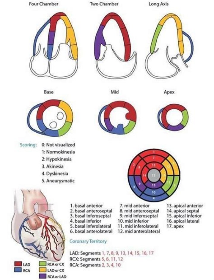
An anterior RWMA (LAD distribution) should correlate with ECG changes in V1 and V2. A lateral RWMA (Cx distribution) should correlate with ECG changes in V5, V6, I, and aVL. An inferior RWMA (RCA distribution) should correlate with ECG changes in II, III, and aVF.
What is a normal LVEF?
The left ventricle is the heart’s main pumping chamber. It pumps oxygen-rich blood up into your body’s main artery (aorta) to the rest of the body. A normal ejection fraction is about 50% to 75%, according to the American Heart Association. A borderline ejection fraction can range between 41% and 50%.
What is RWMA in left ventricle?
Left ventricular RWMA is described as a hypokinesis, dyskinesis, or akinesis of a segment when compared to the other contracting segments of the chamber. This can be visualized sonographically as a blunting of the typical symmetric myocardial thickening during contraction as compared to other cardiac wall segments.
What is PCI stent placement?
Percutaneous Coronary Intervention (PCI, formerly known as angioplasty with stent) is a non-surgical procedure that uses a catheter (a thin flexible tube) to place a small structure called a stent to open up blood vessels in the heart that have been narrowed by plaque buildup, a condition known as atherosclerosis.
Is an echocardiogram serious?
A standard echocardiogram is painless, safe, and does not expose you to radiation. If the test doesn’t show enough images of your heart, though, your doctor might order another procedure, called a transesophageal echocardiogram (TEE).
Can echo detect heart blockage?
Your doctor might recommend a stress echocardiogram to check for coronary artery problems. However, an echocardiogram can’t provide information about any blockages in the heart’s arteries.
What does regional wall abnormalities mean?
Regional wall motion abnormalities are defined as regional abnormalities in contractile function. Ischemic heart disease is the most common cause of wall motion abnormalities. Assessment of wall motion abnormalities is particularly important in the setting of chronic or acute coronary artery disease.