What is rhodopsin cycle?
What is rhodopsin cycle?
rhodopsin, also called visual purple, pigment-containing sensory protein that converts light into an electrical signal. Bleaching and the subsequent regeneration of rhodopsin are major steps in the visual cycle—the series of biochemical reactions that is critical for vision in low light.
What is the function of rhodopsin?
Rhodopsin is a G-protein coupled receptor, and is the most abundant protein in the rod cells found in the retina (Figure 1). It functions as the primary photoreceptor molecule of vision, and contains two parts: an opsin molecule linked to a chromophore, 11-cis-retinal (Athanasiou et al., 2018).
How is rhodopsin activated?
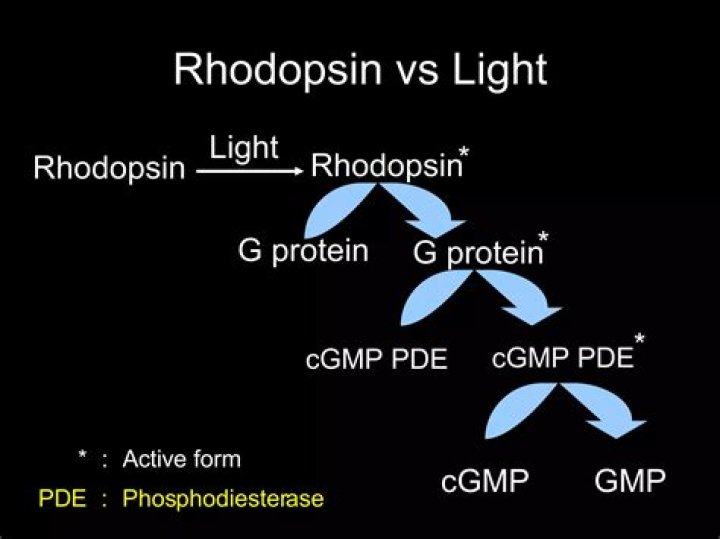
When rhodopsin is activated by light the protein couples with the G protein transducin which is the first step in the signal cascade. Rhodopsin must undergo several conformational changes before being able to bind transducin. Rhodopsin is initially converted to metarhodopsin II which is the active form of rhodopsin.
What is walds visual cycle?
Visual phototransduction is the sensory transduction of the visual system. It is a process by which light is converted into electrical signals in the rod cells, cone cells and photosensitive ganglion cells of the retina of the eye. It is so called “Wald’s Visual Cycle” after him.
What kind of receptor is rhodopsin?
G-protein-coupled receptor
Rhodopsin is a biological pigment found in the rods of the retina and is a G-protein-coupled receptor (GPCR). It belongs to a group of photoswitchable opsins. Rhodopsin is extremely sensitive to light, and thus enables vision in low-light conditions.
Which vitamin is present in rhodopsin?
Rhodopsin is a pigment present in the rod cells of the retina.It consists of two components-opsin and retinal (=retinene)….Question : Mark the vitamin present in rhodopsin.
| Question | Mark the vitamin present in rhodopsin |
|---|---|
| Chapter Name | Neural Control And Coordination |
| Subject | Biology (more Questions) |
| Class | 11th |
What are rods cones?
Rods and cones are the receptors in the retina responsible for your sense of sight. They are the part of the eye responsible for converting the light that enters your eye into electrical signals that can be decoded by the vision-processing center of the brain. Cones are responsible for color vision.
What is macula and fovea?
The macula is the pigmented part of the retina located in the very center of the retina. In the center of the macula is the fovea, perhaps the most important part of the eye. The fovea is the area of best visual acuity. It contains a large amount of cones—nerve cells that are photoreceptors with high acuity.
What is another name for fovea?
Also called the central fovea or fovea centralis. The word “fovea” is the Latin word for “small pit.” The fovea is literally a small depression (in the retina).
What is the difference between opsin and rhodopsin?
As nouns the difference between rhodopsin and opsin is that rhodopsin is (biochemistry) a light-sensitive pigment in the rod cells of the retina; it consists of an opsin protein bound to the carotenoid retinal while opsin is (biochemistry) any of a group of light-sensitive proteins in the retina.