What is Immunolabeling used for?
What is Immunolabeling used for?
Immunolabeling is a biochemical process that enables the detection and localization of an antigen to a particular site within a cell, tissue, or organ.
What is immunostaining used for?
Immunostaining is used in cell biology to study differential protein expression, localization and distribution at the tissue, cellular, and subcellular level.
What is immunofluorescence labeling?
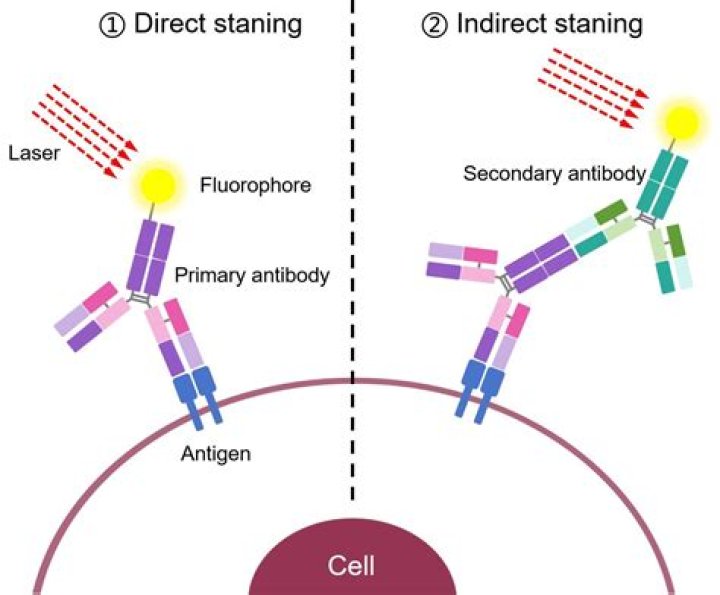
Immunofluorescence is a technique for fluorescently labeling a specific biological target within a sample using an antibody. An antibody is a Y-shaped high–molecular weight glycoprotein, also called an immunoglobulin, that binds specifically (but noncovalently) to another molecule (often called the antigen or epitope).
What microscope is used for immunofluorescence?
Several microscope designs can be used for analysis of immunofluorescence samples; the simplest is the epifluorescence microscope, and the confocal microscope is also widely used. Various super-resolution microscope designs that are capable of much higher resolution can also be used.
What is immunocytochemical staining?
Immunocytochemistry is a technique used to assess the presence of a specific protein or antigen in cells (cultured cells, cell suspensions) by use of a specific antibody, which binds to it, thereby allowing visualization and examination under a microscope.
What is immunofluorescence and its types?
Direct immunofluorescence (DIF) is a one-step histological staining procedure in which tissue antigens (fixed in a solid phase, mostly slides) can be recognized directly by adding fluorochrome-labeled antibodies. From: Reference Module in Biomedical Sciences, 2021.
Where is IHC used?
Immunohistochemistry (IHC) is an important application of monoclonal as well as polyclonal antibodies to determine the tissue distribution of an antigen of interest in health and disease. IHC is widely used for diagnosis of cancers; specific tumor antigens are expressed de novo or up-regulated in certain cancers.
What are the types of immunofluorescence assay?
There are two classes of immunofluorescence techniques, primary (or direct) and secondary (or indirect).
How do you perform immunofluorescence?
All incubation steps take place at room temperature.
- Wash the cells twice and use tweezers to carefully place the coverslip with upturned cells into the humidified chamber.
- Fix with 4 % formaldehyde for 10 minutes and wash 3 ×.
- Permeabilize with 0.1 % TX-100/PBS for 15–20 minutes and wash 3 ×.