The absence of 'a' waves may be seen in atrial fibrillation. An elevated JVP is the classic sign of venous hypertension (e.g. right-sided heart failure). JVP elevation can be visualized as jugular venous distension, whereby the JVP is visualized at a level of the neck that is higher than normal..
Also, what causes increased jugular venous pressure?
Abstract. The internal jugular vein is observed to assess central venous pressure. The most common cause of raised JVP is congestive cardiac failure, in which the raised venous pressure reflects right ventricular failure (Epstein et al, 2003).
Secondly, is seeing JVP normal? Normally only the a and v waves are visible. Conditions associated with an elevated JVP include congestive heart failure and fluid overload.
People also ask, what is jugular vein distention a sign of?
The blood flow from the head to the heart is measured by central venous pressure or CVP. Jugular vein distention or JVD is when the increased pressure of the superior vena cava causes the jugular vein to bulge, making it most visible on the right side of a person's neck.
What causes left jugular vein distention?
Common causes of jugular vein distention Congestive heart failure (deterioration of the heart's ability to pump blood) Hypervolemia (increased blood volume) Superior vena cava obstruction (blockage of the main vein of the upper body that returns blood to the heart; the jugular veins empty into this vein)
Related Question Answers
How do you test for jugular venous distention?
To help determine your CVP, your doctor will actually measure the height of the bulge. While you're lying down on an exam table, with the head of the table at a 45-degree angle and your head turned to the side, your doctor will measure the highest point at which pulsations can be detected in your internal jugular vein.How do you measure jugular venous pressure?
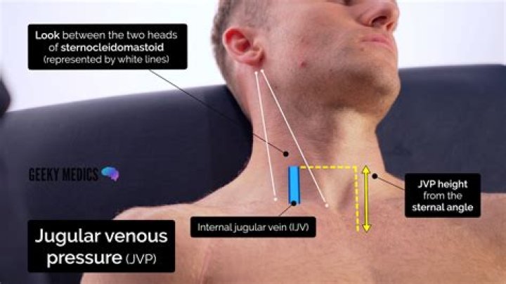
Techniques: Jugular Venous Pressure Measurement (JVP) Neck should not be sharply flexed. Using a centimeter ruler, measure the vertical distance between the angle of Louis (manubrio sternal joint) and the highest level of jugular vein pulsation. A straight edge intersecting the ruler at a right angle may be helpful.Why is my neck vein pulsating?
Abnormal pulses in a woman's neck were caused by a heart valve problem. This causes the right atrium to get bigger, and can change the pressure in nearby blood vessels, potentially leading to abnormal pulses seen in the neck veins, according to the American Heart Association.What is normal venous pressure?
Central venous pressure is usually reported as cm H2O (1 cm H2O = 0.736 mmHg). Normal central venous pressure ranges from 0 to 5 cm H2O. Pressures above 12 cm H2O might indicate hypervolemia or cardiac failure.Why is the jugular vein so important?
IT'S A REAL BRAIN DRAIN. "The jugular vein is an important body part because it drains deoxygenated blood from the head and the neck," Ashley tells Mental Floss. "Most important is to drain the brain. If you block the jugular veins, the pressure in the brain goes up."What is Kussmaul sign?
Kussmaul sign is a paradoxical rise in jugular venous pressure (JVP) on inspiration, or a failure in the appropriate fall of the JVP with inspiration. It can be seen in some forms of heart disease and is usually indicative of limited right ventricular filling due to right heart dysfunction.What is the most common cause of jugular venous distention JVD?
JVD is often caused by life-threatening conditions such as pulmonary embolism, tension pneumothorax, car- diac tamponade, and heart failure,1 and is a classic and crucial finding in the evaluation of all patients presenting with shock.Where is your jugular vein located?
They each rest beside the thyroid gland at the center of the neck, just above the collarbone and near the trachea, or windpipe. These veins functions to carry oxygen-depleted blood from the brain, face, and neck, and transport it to the heart through the superior vena cava.How is venous pressure measured?
The central venous pressure is measured by a central venous catheter placed through either the subclavian or internal jugular veins. The central venous pressure can be monitored using a pressure transducer or amplifier. First, the transducer or amplifier must be zeroed to atmospheric pressure.How do I report JVP findings?
Extend card or ruler horizontally from highest pulsation point , cross with ruler placed on the sternal angle (Angle of Louis), (let's say it was 8cm). Add 5 cm (to get to the center of the atrium) and then report the JVP as "the jugular venous pressure was 13 cm of water" (not mercury).What is the function of jugular veins?
The jugular veins are veins that take deoxygenated blood from the head back to the heart via the superior vena cava.Why JVP is seen on the right side of the neck?
The jugular venous pressure is usually assessed by observing the right side of the patient's neck. The c wave is caused either by transmission of the carotid arterial impulse through the external and internal jugular veins or by the bulging of the tricuspid valve into the right atrium in early systole.