What does a subarachnoid hemorrhage look like on CT scan?
What does a subarachnoid hemorrhage look like on CT scan?
On CT scans, subarachnoid hemorrhage (SAH) appears as a high-attenuating, amorphous substance that fills the normally dark, CSF-filled subarachnoid spaces around the brain, as shown in the images below. The normally black subarachnoid cisterns and sulci may appear white in acute hemorrhage.
How do you describe a subarachnoid hemorrhage?
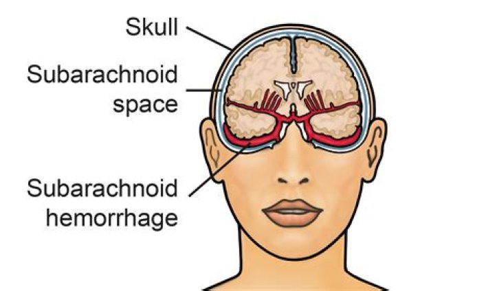
Subarachnoid hemorrhage (SAH) is bleeding into the subarachnoid space. This is usually found centrally (around the circle of Willis) but can occur in other parts of the brain.
Can subarachnoid hemorrhage be missed on CT?
Aneurysmal Subarachnoid Hemorrhage Is Often Missed on Initial Head CT. Retrospective review by neuroradiologists found that CT evidence of SAH was missed in half of CT scans reported to be negative for aSAH.
Does a CT scan show hemorrhage?
Although serial CT scans showed no evidence of hemorrhage, a subacute intracerebral hemorrhage was demonstrated by magnetic resonance imaging.
What are common complications of SAH?
Complications of SAH include the following:
- Hydrocephalus.
- Rebleeding.
- Delayed cerebral ischemia from vasospasm.
- Intracerebral hemorrhage.
- Intraventricular hemorrhage.
- Left ventricular systolic dysfunction.
- Subdural hematoma.
- Seizures.
How are SAH graded?
The Fisher grading system is used to classify SAH, as follows: Grade 1 – No subarachnoid blood seen on CT scan. Grade 2 – Diffuse or vertical layers of SAH less than 1 mm thick. Grade 3 – Diffuse clot and/or vertical layer greater than 1 mm thick.
What shows up as white on a CT scan?
A special dye called contrast material is needed for some CT scans to help highlight the areas of your body being examined. The contrast material blocks X-rays and appears white on images, which can help emphasize blood vessels, intestines or other structures.
What does acute hemorrhage look like on CT scan?
Acute hematoma is seen by pre-contrast CT imaging as an area of high density. CT can detect acute intracerebral blood as small as 2 mm, due to contrast between high density of blood and low density of surrounding brain. Etiology for intracranial hemorrhage: Trauma.