What causes parotid duct obstruction?
What causes parotid duct obstruction?
Parotid duct obstruction is most often caused by salivary gland stones. These are tiny stones made of calcium and other minerals. You’re more likely to have salivary gland stones if you: Have an infection in the parotid gland.
What is duct of Stensen?
The parotid duct, or Stensen duct, is the major duct of the parotid gland, which is the major salivary gland. This duct serves as a conduit for saliva between the substance of the parotid gland and the oral cavity.
What causes buccal mucosa?
Buccal mucosa is the lining of the cheeks and the back of the lips, inside where they touch the teeth. Development of cancer cells or tumour in this area leads to carcinoma buccal mucosa, which is a type of oral cancer. It generally starts in the squamous cells that are thin and flat, and line the lips and the mouth.
Is buccal mucosa a cancer?
Inner cheek cancer (also called buccal mucosa cancer) is a type of head and neck cancer that begins when the cells that make up the inner cheek grow out of control and form lesions or tumors. Buccal mucosa is another name for the inside lining of the cheeks.
How is parotid duct obstruction treated?
How is parotid duct obstruction treated?
- Increasing fluids.
- Putting moist heat on the area.
- Massaging the gland and duct.
- Sucking on candies to promote saliva secretion.
- Using pain medicines.
- Stopping use of any medicines that decrease saliva production, if medically possible.
Do parotid cysts have to be removed?
Because parotid cysts continue to grow over time and are prone to infection, it is important to have them surgically removed to prevent long-term complications.
Why is it called Stensen duct?
It is named after the Danish anatomist Niels Stensen (1638-1686) 2 (also known as Nicolaus Steno) who was the first to describe it, initially in a sheep, in 1660. His colleague Sylvius (1614-1672) confirmed its presence in the human body and van Horne in Leyden named it after Stensen 6.
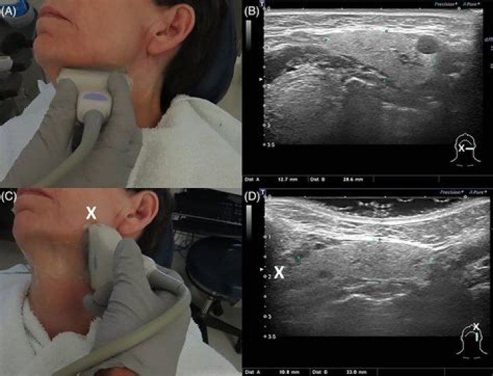
How do you examine a Stensen duct?
Perform a flow assessment at Stensen’s ducts by first positioning your light correctly, pulling the buccal mucosa out to expose the duct, and then drying all fluid from the buccal mucosa area. Next, compress the parotid gland externally with the other hand moving from posterior to anterior across the gland.
What is a buccal lesion?
The buccal mucosa at the occlusal line (cheek-biting), lower lip vestibule, lateral tongue and edentulous ridges (where mastication of food makes contact with the ridge) are common sites. The lesion is often slightly textured and white, and takes on the shape or outline of the traumatic cause (Figure 9).