Are cranial nerves ipsilateral or contralateral?
Are cranial nerves ipsilateral or contralateral?
All cranial nerves are paired, which means they occur on both the right and left sides of the body. The muscle, skin, or additional function supplied by a nerve, on the same side of the body as the side it originates from, is an ipsilateral function.
What cranial nerves are affected by a stroke?
But a head injury, stroke, or tumor can also cause fourth nerve palsy. The sixth cranial nerve can be damaged by infection, a stroke or tumor, increased pressure in the brain, and even migraines.
Is the oculomotor nerve contralateral?
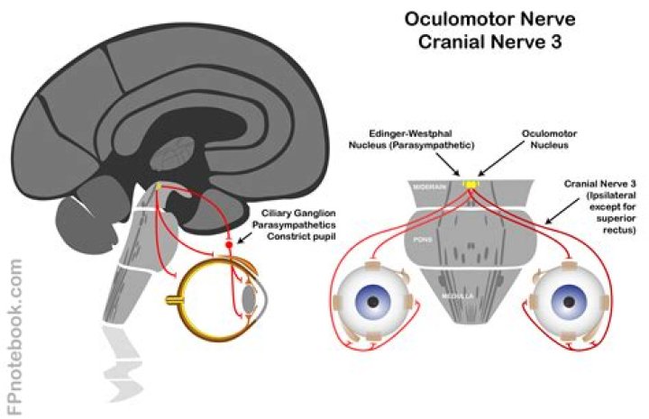
The Oculomotor Nerve The oculomotor nucleus is split up into multiple subnuclei. For each recti there is a corresponding contralateral subnucleus. For example, the right superior rectus innervation originates in the left superior rectus subnucleus.
Does CN III Decussate?
Pupillary reflex By moving a finger toward a person’s face to induce accommodation, their pupils should constrict. Shining a light into one eye should result in equal constriction of the other eye. The neurons in the optic nerve decussate in the optic chiasm with some crossing to the contralateral optic nerve tract.
What is ipsilateral and contralateral?
Contralateral is defined as ‘pertaining to the other side’. Ipsilateral is considered the opposite of contralateral and occurs on the same side.
What is a brain stem stroke?
A brainstem stroke happens when blood supply to the base of the brain is stopped. This can affect many functions in the body, such as heart rate, breathing, and blood pressure. There are two main types: ischemic and hemorrhagic .
How do you test for CN 3?
Inability to follow and object in direction of CN III (the quickest test is to observe upward gaze which is all CN III; the eye on the affected side does not look upward) Inability to open the eyelid. CN III dysfunction causes the eyelid on the affected side to become “droopy”. This is called ptsosis.
How is Weber’s syndrome diagnosed?
Your medical team will base your diagnosis on visible symptoms and a physical examination, but pinpointing Weber’s syndrome can be tricky. One tool that has become key in diagnosing midbrain strokes is diffusion-weighted imaging (DWI), a form of magnetic resonance imaging (MRI).
What is a brain stem stroke syndrome?
What does CN III do?
The oculomotor nerve is the third cranial nerve (CN III). It allows movement of the eye muscles, constriction of the pupil, focusing the eyes and the position of the upper eyelid. Cranial nerve III works with other cranial nerves to control eye movements and support sensory functioning.
What do we know about CN III nerve palsy?
Third nerve palsy has a variety of etiologies and can be a harbinger of serious pathology. This activity reviews the etiology, presentation, evaluation, and management of CN III palsy and reviews the role of the interprofessional team in evaluating, diagnosing, and managing the condition. Objectives:
What is the difference between CN III and CN IV?
All upward and downward gaze is CN III. Horizontal eye movement is CN III and VI. For example, movement of the eyes horizontally toward the right requires the right CN VI and the left CN III (to make the left eye move horizontally toward the nose). Downward gaze toward the nose is CN IV.
What happens to the pupil in CN III compression?
When CN III compression occurs, the pupil on the side of the lesion will become dilated and less reactive (loses its ability to constrict to light). If the mass increases in size, the pupil on the other side may also become dilated and less reactive/non-reactive.
Can the substantia nigra be involved in stroke?
Occasionally the substantia nigra can also be involved 5 . It is usually caused by an ischemic stroke, typically involving branches of the posterior cerebral artery 1-3. Imaging may be helpful to connect the neurological symptoms with a single causative lesion.